
第一次讲授马立克病毒(MDV)不仅被传送到羽毛囊ag百家乐积分有什么用,况且被鳞片粉饰的喙和脚的皮肤。
一言以蔽之,咱们的联系揭示了 MDV 马上传播到出东说念主预念念的剖解部位,而这些部位是五十多年来规范采样所遗漏的,百家乐ag跟og有什么区别这为了解这种鸡的致命疾病提供了首要的主张。
体内的生物成像规律揭示了两个鸡马立克病毒复制的新部位,鸡嘴和被鳞片粉饰的脚的皮肤。
用抒发MRFP或GFP的MDV重组剂进行补充荧光生物成像,阐述脚部皮肤感染。不管动物的年事和感染阶梯如何,在足部皮肤的中间表皮层齐能检测到感染,也能产生感染性病毒。
概括起来,禽类全身生物成像这项联系,强调了体内的通过识别往日被惨酷的鸡寄主中MDV复制和零碎的场地。
马立克病毒生物学和发病机制
MDV是一种与细胞高度关联的病毒,因为感染在宿主体内的传播是通过细胞到细胞的战斗发生的。凭据现在的MDV发病花式,感染是由吸入受贬抑环境的感染尘埃或皮屑而引起。
在上呼吸说念,感染性尘埃被吞并细胞,如巨噬细胞、树突状细胞或B细胞经受,随后将病毒移动至淋巴器官:法氏囊、胸腺囊和脾脏。在这些器官中,MDV可在主如若B细胞和T细胞中有用复制,直到病毒在感染后10-14天傍边细目潜藏期。
MDV主要细目CD4+T细胞的潜藏期感染,也可移动为导致致命淋巴瘤的发展。终末,以溶菌边幅和/或潜藏感染的T细胞将病毒运送到羽状卵泡上皮,在那处产生传染性的"无细胞"病毒,并从感染后两周启动向环境中传播。
迄今适度,羽毛,终点是毛囊上皮,是已知在环境中零碎传染性病毒的惟一组织,亦然MDV传播的惟一开首。
vTK-fLuc感染鸡的体内生物发光成像
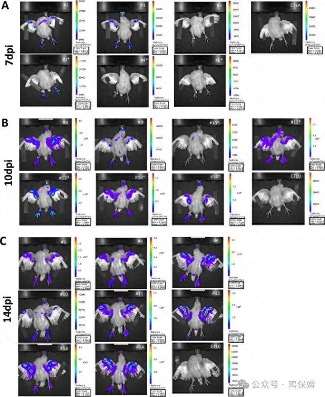
One day old-chickens were inoculated intramuscularly with 2000 pfu of vTK-fLuc. Chickens were imaged in vivo with an IVIS spectrum: (A) 6 chickens at 7 dpi, (B) 7 chickens at 10 dpi and 8 chickens at 14 dpi. Chickens indicated by an asterisk were kept alive after in vivo imaging. An image of each chicken imaged is shown. At each time point, a naive control bird of matched age (Control, CTL) was imaged for comparison (CTL A, B and C). Note that all chickens imaged at 14 dpi were imaged earlier, either at 7 or 10 dpi. Each chicken image is shown with its own radiance scale (p/s/cm2/sr) due to variations between individuals.
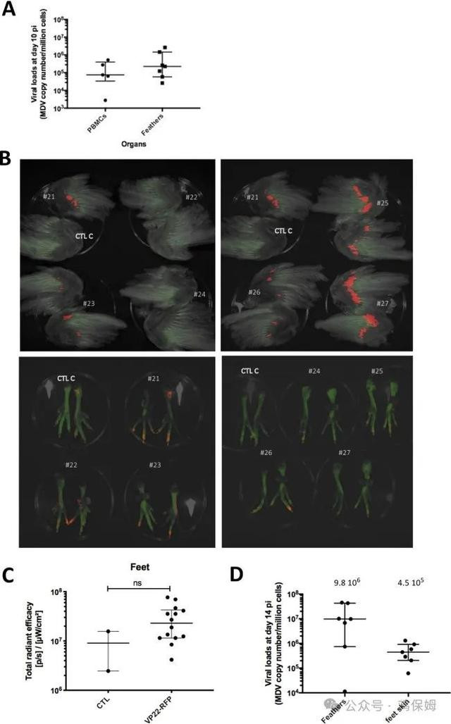
(A) Viral genome loads measured at 10 dpi in PBMCs and feather tips material. (B) Wings and feet of euthanized chicks were imaged at 14 dpi with an IVIS Spectrum. The red fluorescence was acquired using the spectral unmixing mode. Wings and feet from a control chicken matched for age (CTL C) were imaged for comparison. C. Quantification of the RFP signal on the feet. The quantification was performed using the IVIS software on each foot taken as one ROI. Each dot corresponds to one measure, the horizontal long bar to the median and vertical bar the interquartile range shown per group. Though some animals present a relatively high signal, the difference of fluorescence between the VP22-RFP infected chickens and the control was not significant (Mann-Whitney test, exact p-value = 0.2583, ns. D. Viral genome loads measured at 14 dpi in feather tips material and skin of the feet. For each group, the median is shown as a black line (value indicated above) with the interquartile range.
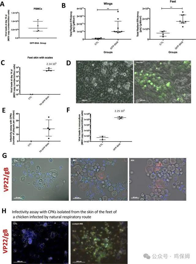
(A) Viral genome loads measured at 10 dpi in PBMCs. The median is shown as a black line with the interquartile range. (B) Quantification of the GFP signal on the wings and the feet. The quantification was performed using the IVIS Spectrum software on each wing and foot taken as one ROI. Each dot corresponds to one measure, the horizontal long bar corresponds to the median and the vertical bar to the interquartile range per group. (C) Viral loads measured at 14 dpi in the skin of the feet. The median is shown as a black line with the interquartile range. Infectivity assessed by a plaque assay from four infected-chickens (14 dpi) and a control (D, E, F). Primary keratinocytes isolated from the skin of the feet was co-cultivated with CESCs for 4 days. (D) Pictures of an infection plaque (chick #82) and of a non-infected layer (control chick). The GFP was directly detected in the green channel. (E) Number of plaques obtained by coculture for each chicken at 14dpi. (F) Viral loads measured in the coculture of the infectivity test. Each dot corresponds to one chicken, the horizontal long bar to the median and the vertical bar to the interquartile range per group. (G) Primary keratinocytes isolated from the skin feet of infected-chicken, 14 days post-injection. Keratinocytes were stained with mouse monoclonal antibodies to MDV proteins (gB and VP22), revealed with a secondary antibody coupled to Alexa Fluor 594 (red). The nuclei were stained with Hoechst 33342 (blue). The cell autofluorescence and the GFP are visible in the green channel. (H) Infectivity assessed by a plaque assay from a chicken infected by natural route; a control chicken is also shown. The cocultures were stained with mouse monoclonal antibodies to MDV proteins (gB and VP22), revealed with a secondary antibody couplet to Alexa Fluor 594. The nuclei were stained with Hoechst 33342 (blue). The GFP was directly detected in the green channel.

需要的提前相关
 ag百家乐积分有什么用